Quantcast
ZME Science
  • News
  • Environment
  • Health
  • Future
  • Space
  • Features
  • ZME & more
    • About
    • The Team
    • Advertise
    • Contribute
    • Our stance on climate change
    • Privacy Policy
    • Contact
No Result
View All Result
ZME Science

No Result
View All Result
ZME Science
Home Health & Medicine Anatomy

Scientists image organs at microscopic scales

In a new study published in Nature, researchers have demonstrated a technique that allows the mapping of organs at microscopic scales.

by Mihai Andrei
November 26, 2015
in Anatomy, News, Physics
A A

In a new study published in Nature, researchers have demonstrated a technique that allows the mapping of organs at microscopic scales; they detail its use and produced images of the microvessels in the brain of a live rat.

Image of the whole brain vasculature at microscopic resolution in the live rat using ultrafast Ultrasound Localization Microscopy: Local density of intravascular microbubbles in the right hemisphere, quantitative estimation of blood flow speed in the left hemisphere. Credit: ESPCI/INSERM/CNRS
Image of the whole brain vasculature at microscopic resolution in the live rat using ultrafast Ultrasound Localization Microscopy: Local density of intravascular microbubbles in the right hemisphere, quantitative estimation of blood flow speed in the left hemisphere.
Credit: ESPCI/INSERM/CNRS

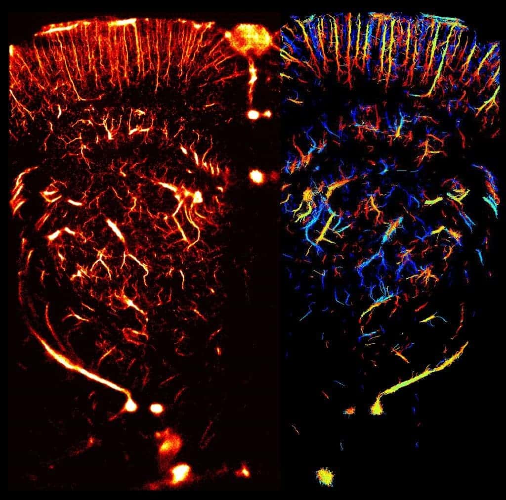
Current techniques of microscopic imaging are not ideal. Just like in geological imaging techniques, there’s a tradeoff between penetration depth, resolution, and time of acquisition. In other words, if you want to see all the way through the human body, you won’t have good enough resolution – or you will but it will take a very long time. Until now, conventional imaging techniques have worked at milimetre and sub-milimetre scale at best, being limited by one of the fundamental laws of physics: features smaller than the wavelength of the radiation used for imaging cannot be resolved. But Mickael Tanter, a professor at the Langevin Institute and his colleagues have come up with a new approach, and report a new super-resolution.

Like a few other recent studies, they used microbubbles, with diameters of 1–3 micrometres, which were injected into the bloodstream of live rats. They then combined deep penetration and super-resolution imaging in a technique they call ‘ultrafast ultrasound localization microscopy’. With this, they obtained ultra fast frame rates (500 / second), and achieved a resolution equal to the rat brain microvasculature – less than 10 micrometers in diameter (0.01 milimetres).

While this is still a work in progress, authors believe it can help doctors make better diagnostics and better understand how some diseases affect the body.


Tags: bodyimagingorgan

ADVERTISEMENT
ADVERTISEMENT
  • News
  • Environment
  • Health
  • Future
  • Space
  • Features
  • ZME & more

© 2007-2021 ZME Science - Not exactly rocket science. All Rights Reserved.

No Result
View All Result
  • News
  • Environment
  • Health
  • Future
  • Space
  • Features
  • ZME & more
    • About
    • The Team
    • Advertise
    • Contribute
    • Our stance on climate change
    • Privacy Policy
    • Contact

© 2007-2021 ZME Science - Not exactly rocket science. All Rights Reserved.