Quantcast
ZME Science
  • News
  • Environment
  • Health
  • Future
  • Space
  • Features
  • More
    • About
    • The Team
    • Advertise
    • Contribute
    • Our stance on climate change
    • Privacy Policy
    • Contact
No Result
View All Result
ZME Science

No Result
View All Result
ZME Science
No Result
View All Result
Home Environment Animals

Laser-light sheets used to image life at its earliest stage [GREAT PICS]

Tibi Puiu by Tibi Puiu
June 10, 2013
in Animals, Biology, Great Pics
Reading Time: 1 min read
A A
Share on FacebookShare on TwitterSubmit to Reddit
Image of the ~50,000 cell nuclei of a 22-hour-old zebrafish embryo. The fluorescently labeled cell nuclei are shown in a blue-to-red color code that indicates depth in the image.

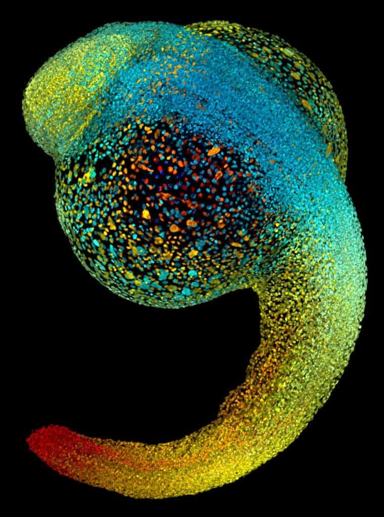
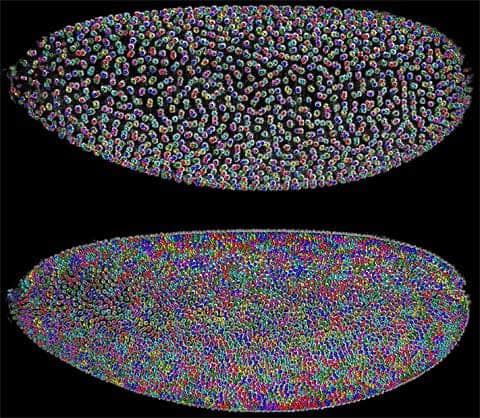
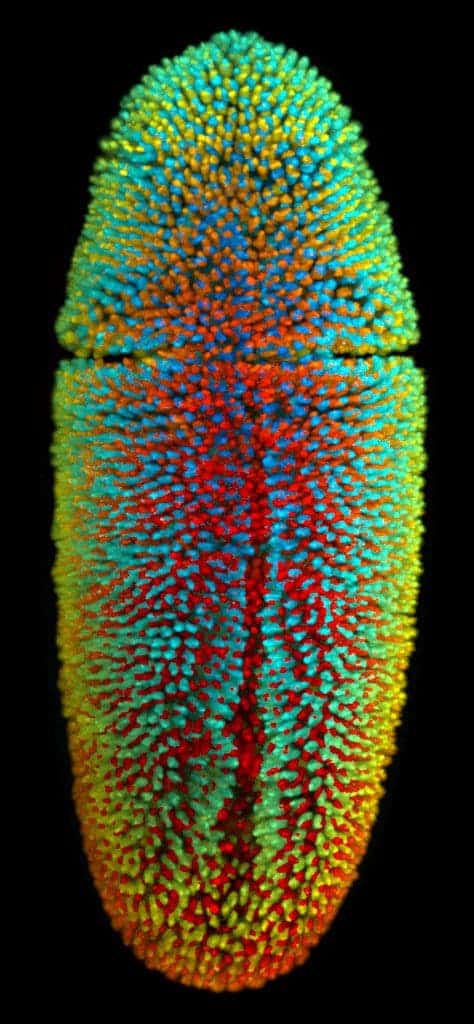
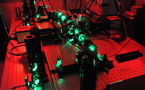
A new visualization technique developed by researchers at the Howard Hughes Medical Institute used a thin sheet of laser light that beams, stepwise, into different planes of a specimen to create intricate and detailed snapshots of cells. In these pictures featured above and below you can see how zebrafish and fruit fly embryos were imaged using this novel technique.

By following the color-coded cells of a Drosophila embryo (top) over time, each cell’s lineage becomes trackable (bottom) with simultaneous multi-view light sheet microscopy.
Image of the ~6,000 cell nuclei of a 3-hour-old fruit fly embryo. The fluorescently labeled cell nuclei are shown in a blue-to-red color code that indicates depth in the image.

Here’s how it all works:

Sorry to interrupt, but you should really...

...Join the ZME newsletter for amazing science news, features, and exclusive scoops. More than 40,000 subscribers can't be wrong.


“The laser light causes the cells in the illuminated plane to fluoresce while a set of two or four cameras gather snapshots of every cell in the plane from several different angles. By taking pictures as the embryo rotates through the beam, Keller collects a set of planar views which are assembled into a dynamic three-dimensional depiction of the embryo at any given time during its 21 or more hours of development.”

A SiMView microscope uses lasers to illuminate specimens while two cameras to the left and to the right of the central imaging chamber capture shots of the specimen’s cells from different angles.
ADVERTISEMENT
ADVERTISEMENT
A SiMView microscope uses lasers to illuminate specimens while two cameras to the left and to the right of the central imaging chamber capture shots of the specimen’s cells from different angles.

Tags: embryo
ShareTweetShare
Tibi Puiu

Tibi Puiu

Tibi is a science journalist and co-founder of ZME Science. He writes mainly about emerging tech, physics, climate, and space. In his spare time, Tibi likes to make weird music on his computer and groom felines.

ADVERTISEMENT
ADVERTISEMENT
  • News
  • Environment
  • Health
  • Future
  • Space
  • Features
  • More

© 2007-2019 ZME Science - Not exactly rocket science. All Rights Reserved.

No Result
View All Result
  • News
  • Environment
  • Health
  • Future
  • Space
  • Features
  • More
    • About
    • The Team
    • Advertise
    • Contribute
    • Our stance on climate change
    • Privacy Policy
    • Contact

© 2007-2019 ZME Science - Not exactly rocket science. All Rights Reserved.