These aren’t Christmas lights, but the actual neural activity of Caenorhabditis Elegans, a parasitic nematode. The brain imaging was done by researchers at Princeton University, and no worm had to be cut open. Instead, the researchers used a special protein which fluoresces in response to calcium.
When scientists tap the brain, they’re looking for one prime indicator: electrical activity. When a neuron is active, it fires an action potential which is basically a depolarization made between a neuron’s axon to another neuron it signals to. Now, traditionally neuroscientists use a technique called electrophysiology to study the patterns of neuron electrical activity. It’s precise, yet the analysis is limited to a handful of neurons at a time. A more interesting method exploits the fact that when a neuron is active (again, depolarized), calcium flows into it. Using special dyes (proteins) that fluoresce in response to whether or not they bind to calcium, scientists can monitor these calcium dynamics and in turn the depolarization.
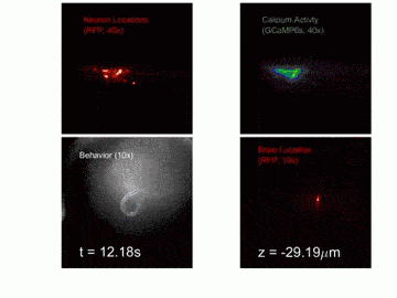
That’s exactly what the Princeton researchers achieved, allowing them to monitor in real time 77 of the nematode’s 302 neurons as they light up. These have been shared in this amazing video, split into four frames. In the upper left, we see the location of the neurons, while the upper right shows a simulation of the calcium signaling which is analogous to neural electrical patterns. In the lower two panels we zoom out: the worm itself (left) and the location of the brain (right).
Using this data, the researchers would like to devise a mathematical model that will allow them to simulate and control the worm’s brain. Previously, other efforts identified how C. elegans can identify magnetic fields, while a more ambitious team from Harvard targeted laser pulses at the worm’s neurons, and directed it to move in any directions they wanted, even tricking the worm in thinking there’s food nearby.
Was this helpful?


