Researchers took a nanoscale snap of a living cell membrane for the first time in history
They had to develop a whole new method to do it.
Researchers have snapped the most detailed ever image of a cell membrane, all the way down to the nanoscale. The image could finally settle a long-standing debate in biology on how it functions, and introduce a powerful new tool to biologists’ toolkit.
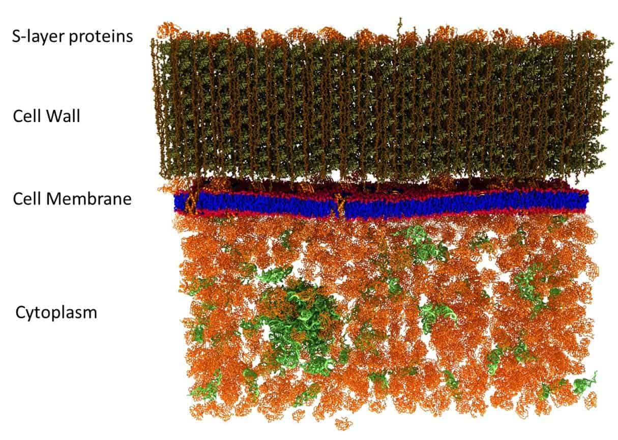
Image credits John Katsaras et al, PLOS ONE (2017).
Every living cell’s membrane is put together from a thin sandwich of lipids, which are fat molecules, interspersed with other bits of organic materials such as proteins and carbohydrates. It’s a pretty nifty system — the fatty bilayer, for example, keeps all the watery stuff inside the cell from mixing with the watery stuff outside the cell. Proteins act as pumps and decide what goes through the membrane and when, or serve as landing areas for signaling molecules so the cell can talk with its pals. Some carbohydrates act as ID tags. Then there’s one other bit whose function — as far as can be summarized if you keep tabs on cytological debates, which I’m sure most of you do — seems to be solely to sow discord and disagreement into the ranks of biologists.
These tiny bits are known as lipid rafts and, although there’s a pretty solid body of documentation as to what they are and what they do, haven’t really caught with all cellular biologists. The short of it is that they act as independent, more compact domains than the rest of the membrane, making it behave a little wobbly, and their movements allow the cell to activate or inactivate proteins along its membrane.
So, a team led by John Katsaras, Senior Biological Systems Scientist at Oak Ridge National Laboratory’s Neutron Sciences Directorate, decided to take a picture and find out.
“It became a debate,” Katsaras said. “Some people believed they exist, while others believed they didn’t. There was a lot of circumstantial evidence that could support either side.”
The way they went about it could fundamentally change how living nanoscale structures are studied in the future.
Looking at the really small
When biologists want to take a peek at the going-ons inside a cell, they normally use fluorescent compounds designed to attach to a particular molecule and tag it, making it visible under the optical microscope. But since we don’t really know what lipid rafts do (so we don’t know where to add the fluorescent tags), and because they’re probably too tiny to spot under the microscope, this doesn’t really work in their case.

Good luck spotting anything.
Image credits Tobi Luxe.
An electron microscope could probably make them out with ease, but the thing is that to find out how these rafts behave you need to observe a living cell. Since cells are made so tiny, atoms are basically brick-sized compared to them. Electrons, then, are bullet- or pellet-sized. To a living cell, an electron microscope is basically a death-spewing chaingun. So that won’t work either.
In the end, the team decided to use a mix of genetic and chemical labeling techniques to add a hydrogen isotope to the membranes of living Bacillus subtilis cells. Then, they used a method called neutron scattering to chart the arrangement of different molecules in the bacterium’s cell membrane. Neutron scattering was picked because it’s less energetic than electron microscopy, meaning the particles aren’t (necessarily) deadly to the bacteria.
So why are the isotopes there? Well, although less energetic, neutrons are way heavier than electrons. So it’s not exactly deadly, but the particles are powerful enough to affect the cell and interfere with its membrane’s internal processes. Furthermore, while it could spot the rafts, neutron scattering couldn’t tell it apart from the rest of the membrane, so the team needed to tag them with something that stands out.
Bag and tag
Since 99.98% of all hydrogen atoms currently in existence only have a single proton for a nucleus, the isotopes the team used, which have an extra neutron attached to the nucleus and are known as deuterium, is pretty conspicuous. And while they chemically function the same (since neutrons don’t affect the atom’s valence/electrical balance), physically they do differ enough to scatter neutrons in a different way — so they were both easy to spot and unlikely to occur naturally.
The team genetically edited a new strain of B. subtilis with a slightly different ratio of hydrogen to deuterium in its membrane compared to wild strains. If there were no rafts, they should see a uniform distribution of these altered fat molecules throughout the membrane.
Instead, their imaging showed areas with pronounced differences in lipid arrangement, which matched the proposed size of the lipid rafts — very strong evidence for their existence. Even better, the technique they developed for the study could fundamentally change how biologists peer into the workings of living cells.
“The people who study these things tend to use particular types of probes,” says Katsaras.
“They didn’t use neutron scattering because it wasn’t in the biologist’s wheelhouse. Our novel experimental approach opens up new areas of research.”
These differentiated areas aren’t visible in the team’s model, but it does an exemplary job of showing how a cell’s outer layers are structured — watery cytoplasm covered with the lipid layer the team was investigating in the middle, and the outer cell wall at the top.
The full paper “The in vivo structure of biological membranes and evidence for lipid domains” has been published in the journal PLOS ONE.