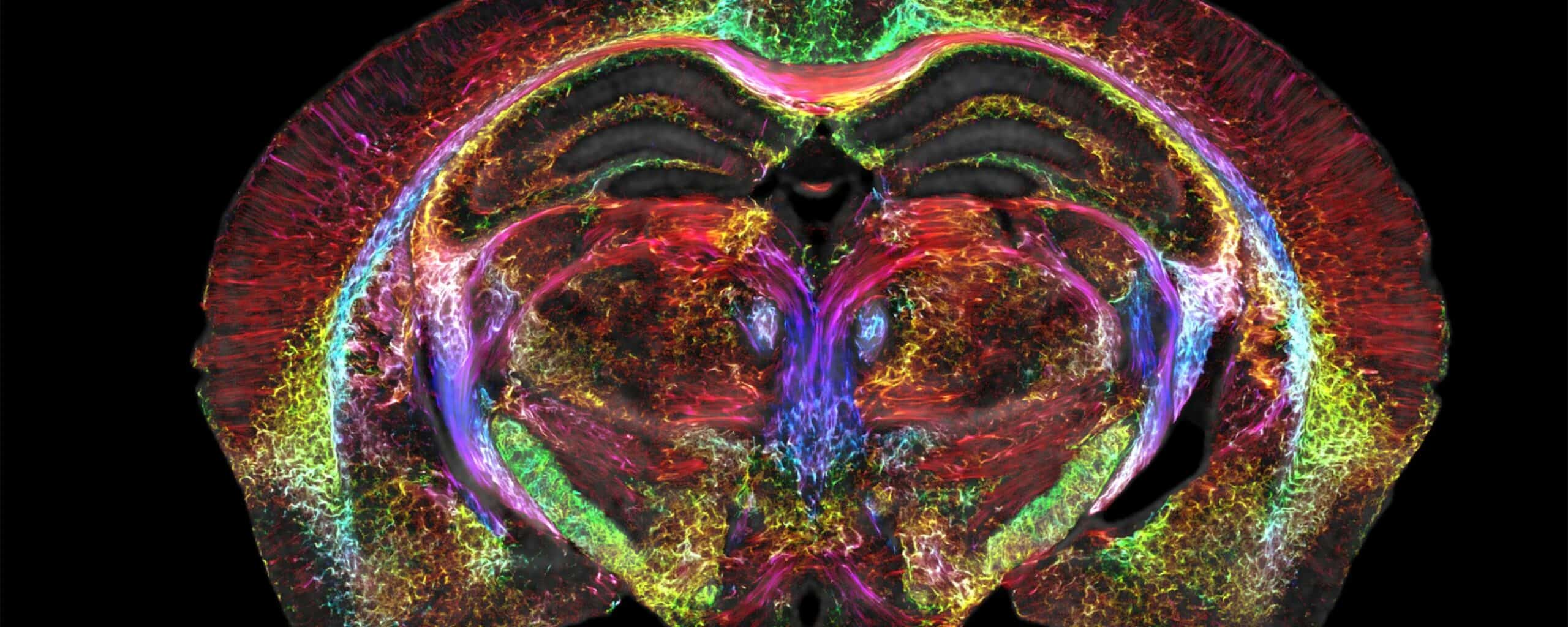
Brain MRIs just got a thousand times sharper
Although the technology was only demonstrated on mice, researchers think it can also work on humans.
“It is something that is truly enabling. We can start looking at neurodegenerative diseases in an entirely different way,” said G. Allan Johnson, the lead author of the new paper.
Magnetic resonance imaging (MRI) has become the ‘workhorse’ of brain imaging, and it’s useful for detecting internal health problems such as bleeding, swelling, tumors, infections, and many other types of damage. In the 50 years since it was invented, the resolution of MRI has been steadily improving, but there’s still plenty of room for progress.
Johnson is part of a group of researchers from several universities that, for decades, has embraced the challenge of improving MRI resolution. They improved and refined the elements that make up MRI machines and in their new breakthrough, they achieved a “resolution which is ~ 1,000 times higher than that of most preclinical MRI”.
MRI works through a mixture of magnets and radio waves. When a patient is placed in the MRI machine, their body is exposed to a strong magnetic field. This causes the protons in the hydrogen atoms within the body to align with the direction of the magnetic field.
Then, a radio frequency pulse is then applied to the patient, which causes the protons to absorb energy and flip their alignment. When the radio frequency pulse is turned off, the protons return to their original alignment and release the energy they absorbed in the form of radio waves.
These radio waves are detected by the MRI machine’s receiver coils and processed by a computer to produce a detailed image of the internal body structure being examined. You can also change the parameters of the magnets and the radio waves to image different types of tissues and different depths.
In the new study, researchers used a special set of coils that are 100 times stronger than those in a clinical MRI. They also deployed massive computing power, the rough equivalent of nearly 800 laptops working together. But the results were worth it.
Just like 2D resolution is measured in pixels (and the smaller the pixel, the better the resolution), 3D resolution is measured in voxels. A voxel in this MRI measures just 5 microns — some clinical MRI voxels are even millions of times larger.
So far, the new technology has only been used on mice and it’s gonna take a bit of extra work to translate it to human MRIs, but even on mice, this could be very useful.
The approach enables researchers to label different types of cells from the brain. For instance, they can label dopamine-producing cells that are related to the progression of Parkinson’s Disease. In fact, mouse models are used extensively in the study of human diseases ranging from Huntington’s disease to Alzheimer’s.
This level of detail could help researchers better understand how things go awry in the mice brains, which in turn, could inform human research.
Indeed, Johnson says that this type of improvement in the level of MRI detail could pave the way for new groundbreaking research in brain diseases and brain-related conditions.
“Research supported by the National Institute of Aging uncovered that modest dietary and drug interventions can lead to animals living 25% longer,” Johnson said. “So, the question is, is their brain still intact during this extended lifespan? Could they still do crossword puzzles? Are they going to be able to do Sudoku even though they’re living 25% longer? And we have the capacity now to look at it. And as we do so, we can translate that directly into the human condition.”
Journal Reference: “Merged Magnetic Resonance And Light Sheet Microscopy Of The Whole Mouse Brain,” G. Allan Johnson, Yuqi Tian, David G. Ashbrook, Gary P. Cofer, James J. Cook, James C. Gee, Adam Hall, Kathryn Hornburg, Yi Qi, Fang-Cheng Yeh, Nian Wang, Leonard E. White, Robert W. Williams. Proceedings of the National Academy of Sciences, April 17, 2023. DOI: 10.1073/pnas.2218617120