Researchers capture first ever images of microglia eating brain synapses
Eating for success!
It’s not just zombies that chow on brains — for the first time, researchers have filmed microglia chowing down on brain synapses.
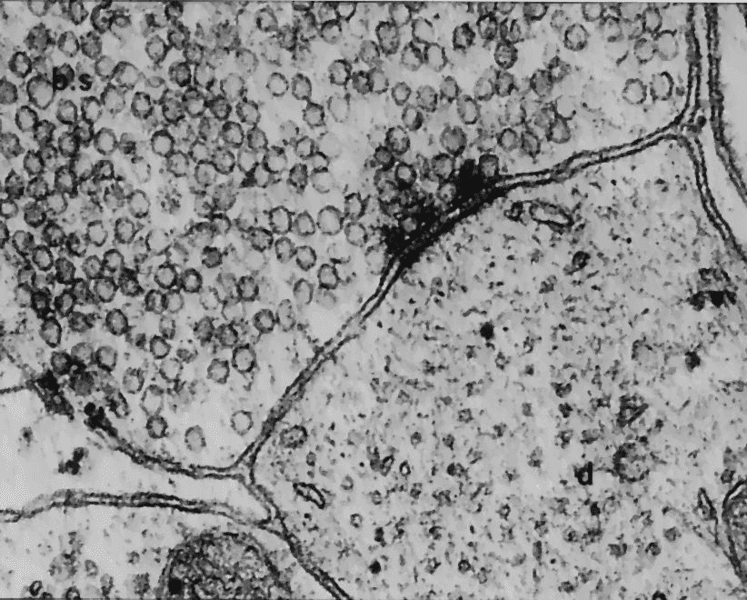
Image credits Savant-fou / Wikimedia.
Brains, like pretty much like your room, tends to get messy. Unlike your room, however, the brain has specialized support cells, a class known as “glia“, to keep everything working — and one type of glia, named “microglia” because they’re quite tiny, act as the brain’s Roombas, keeping it tidy 24/7.
While we knew, in theory, what these microglia were up to, we’ve never actually seen them go about their business. Not until recently, however, when researchers from the European Molecular Biology Laboratory (EMBL) captured them in the act.
“You finished with that?”
Neurons are arguably one of the most important cells in the brain; they’re the ones that process information and perform the job the brain is meant to do. However, the real heavy lifting inside this organ is performed by glia. And they’re quite prolific: our brains are roughly 70% glia, and around 10% of all cells in your brain are microglia specifically.
Microglia are related to macrophages — the variety of white cell that ‘eat’ bad guys. Microglia perform a similar function in the brain, acting as the main active component of its immune system. But they also take an active part in steering brain development; while not fighting anyone, microglia weed out brain synapses that have outlived their usefulness or just to make room for newer, more efficient synapses.
Given their massively influential role in the brain, it’s understandable why researchers have been trying to get a look at microglia in action for a long time now. Researchers from EMBL Rome, led by Laetitia Weinhard, working in collaboration with EMBL Heidelberg, are the first to actually succeed at this task. They set up a massive imaging study to capture the process in action in mouse brains.
The team combined two brain imaging techniques, correlative light and electron microscopy (CLEM), with light sheet fluorescence microscopy — a technique developed at EMBL — to capture the first images of microglia eating synapses.
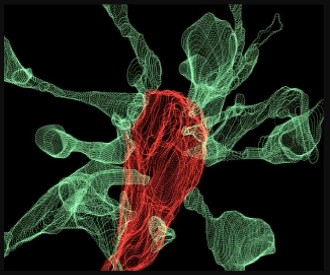
Multiple synapse heads send out filopodia (green) converging on one microglia (red), as seen by focused ion beam scanning electron microscopy. L. Weinhard, EMBL Rome
One surprising finding was that around half of the interactions between microglia and synapses prompted the latter to send out thin projections or ‘filopoda’, as if to greet the cell, the team notes. In one case, the researchers observed no fewer than fifteen synapse heads extending filopoda toward a single microglia as it chowed on another synapse.
“Our findings suggest that microglia are nibbling synapses as a way to make them stronger, rather than weaker,” says Cornelius Gross (EMBL Rome), who led the research.
“As we were trying to see how microglia eliminate synapses, we realised that microglia actually induce their growth most of the time,” Laetitia Weinhard adds.
One other important find is that microglia could underly the formation of double synapses, where one neuron releases neurotransmitters to two others (instead of the traditional one-on-one communication). This mechanism shows that microglia are deeply involved in brain processes such as structural plasticity and can even induce the rearrangement of synapses — a process that underpins learning and memory.
The observations are the product of five years of technological development. During this time, the team worked with three different cutting-edge imaging systems before obtaining the images.
“This is what neuroscientists fantasised about for years, but nobody had ever seen before,” says Cornelius Gross. “These findings allow us to propose a mechanism for the role of microglia in the remodeling and evolution of brain circuits during development.”
Next, the team plans to investigate what role microglia play in brain development during adolescence, or if there’s any link between these cells and mental diseases.


